Endoscopic, Spectroscopic Imaging of the Middle Ear
Contents
Introduction
This project aims to develop a medical device to assess chronic middle ear inflammation in children. For this we collaborate with ENT expert of the Nottingham Hearing Biomedical Research Unit.
Medical Background and Motivation
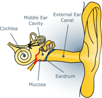
Chronic Middle Ear Inflammation is characterized by inflammation of the middle ear mucosa (marked in red) and secretion of a liquid causing hearing loss. The only effective treatment is surgically draining the liquid and placing a ventilation tube through the eardrum. This is one of the most common surgeries in children in the developed world. In 25% of the cases the symptoms recur and there is no way of predicting the recurrence. It is hypothesised that a recurrence is linked to a persistent inflammation after surgery.

|
This project aims to develop a medical device that will be able to determine whether the middle ear mucosa is still inflamed or has returned to normal in order to improve diagnosis. Direct view of the mucosa is obscured by the eardrum. Consequently, an optical method must be implemented to allow measurements through the eardrum by maximising the signal detected from the mucosa and minimising background reflections from the eardrum. The ratio of the detected signal at two wavelengths will allow assessment of the inflammation similar to haemoglobin concentration measurements. The use of confocal and anti-confocal systems is investigated here.
The Optical System
The optical system aims to solve two problems, first to maximise signal from the mucosa while minimising reflections from the eardrum, and second to spectroscopically assess the middle ear mucosa to determine the state of the inflammation.
Spatial Filtering
Instead of using a confocal system to reject reflected light from the eardrum and select signal from the mucosa an anti-confocal system is introduced. In the anti-confocal system the pinhole is replaced by a stop rejecting light inside rather than outside its radius. This allows to reject reflection from one plane - the eardrum in this case - and detect all other light. This allows a higher signal to be detected and a bigger volume of the mucosa to be sampled.

|

|
Spectroscopic System
The spectroscopic system will make use of the wavelength dependent absorption of haemoglobin in order to measure the blood concentration in the mucosa indicating the metabolism and state of the inflammation. Measurements will be conducted at at least two wavelengths in the visible and near infra-red range.
Progress
Simulations
Imaging of the middle ear using the anti-confocal system has been simulated using Monte-Carlo methods and compared to a conventional confocal system. The simulation shows an improved performance of the anti-confocal system considering signal strength and signal-to-background ratio.
Tissue Phantoms
Animal eardrums have been characterised and investigated in order to find tissue models for the human eardrum. Further, epoxy resin based tissue phantoms have been produced to model healthy and inflamed mucosa. These phantoms will provide a middle ear model for testing of the optical system.
Optical System
The optical system is set up as bench top system. A CCD sensor is used as confocal detector to allow easy variations of the filtering element of the (anti)-confocal system in software during post processing of the measurements.
Future Work
Next steps are to experimentally characterise the optical system and proof the concept using the tissue simulating phantoms. When shown to be working the system can be minimised and adopted for clinical use and tested on human subjects.
Related Publications
- Jung, D.S.; Birchall, J.P.; Crowe, J.A.; See, C.W.; and Somekh, M.G., "Monte Carlo Photon Simulation of an Anti-Confocal System to Monitor Middle Ear Inflammation," in Optics in the Life Sciences, OSA Technical Digest (CD) (Optical Society of America, 2015), paper JT3A.41.
- Jung, D.S.; Crowe, J.A.; Birchall, J.P.; Somekh, M.G.; and See, C.W., "Anti-confocal versus confocal assessment of the middle ear simulated by Monte Carlo methods," Biomed. Opt. Express 6, 3820-3825 (2015).