Difference between revisions of "High frequency ultrasonics using optical fibres"
(Created page with "==Motivation== Optical fibres have been used to propel the fields of optical and acoustic endoscopy, due to their ability to transmit information (such as an image) in and ou...") |
|||
| (3 intermediate revisions by the same user not shown) | |||
| Line 1: | Line 1: | ||
==Motivation== | ==Motivation== | ||
| − | Optical fibres have been used to propel the fields of optical and acoustic endoscopy, due to their ability to transmit information (such as an image) in and out of extremely confined environments. There is great interest in developing an optical fibre probe which functions using GHz frequencies of ultrasound. This would allow high resolution inspection of microscopic environments and specimens, where lateral resolution would be dictated by the diamter of the fibre core (~ 5 μm), and axial resolution would be determined by the acoustic wavelength (~ 300 nm). Such a device could be implemented in a wide range of scenarios, especially ones in which access to the sample plane is limited and ill-suited for traditional bench-top acoustic and photoacoustic microscopy. For example, optical fibre sensors could be readily integrated through the bore of a needle during fine needle aspiration procedures and other biopsies, or in the form of imaging bundles which are already widely utilised in clinical endoscopes. | + | Optical fibres have been used to propel the fields of optical and acoustic endoscopy, due to their ability to transmit information (such as an image) in and out of extremely confined environments. There is great interest in developing an optical fibre probe which functions using GHz frequencies of ultrasound. This would allow high resolution inspection of microscopic environments and specimens, where lateral resolution would be dictated by the diamter of the fibre core (~ 5 μm), and axial resolution would be determined by the acoustic wavelength (~ 300 nm). The fact that the device operates acoustically means that label-free high contrast can be achieved through the mechanical properties of the specimen, while still remaining non-contact. Such a device could be implemented in a wide range of scenarios, especially ones in which access to the sample plane is limited and ill-suited for traditional bench-top acoustic and photoacoustic microscopy. For example, optical fibre sensors could be readily integrated through the bore of a needle during fine needle aspiration procedures and other biopsies, or in the form of imaging bundles which are already widely utilised in clinical endoscopes. |
| − | |||
| − | |||
| − | |||
| − | |||
| − | |||
==Brillouin fibre-spectrometer== | ==Brillouin fibre-spectrometer== | ||
| + | The phonon probe consists of a single mode optical fibre which has been coated with 15 nm of gold onto its distal end. This fibre transducer is then integrated into a pump-probe spectroscopy system consisting of two pulsed lasers with 100 fs pulse widths. The pump and probe pulses are synchronised using asynchronous optical sampling (ASOPS) in order to vary the time delay between the two beams over a time window of ~10 ns. The gold serves as a photoacoustic source which, upon absorption of the pump beam, thermoelastically generates ultrasound within a ~100 GHz bandwidth. When coupled with a liquid medium such as water, the GHz acoustic waves periodically modulate the refractive index of the medium due to the photoelastic effect. This sets up a moving diffraction grating for the probe beam, and results in Brillouin scattering. The interference of the inelastically reflected probe beam with an elastically scattered reference beam modulates the optical reflectance creating a time-resolved reconstruction of the acoustic mode. | ||
| + | {|align="center" | ||
| + | |[[File:Sal_fig1.png|800px|thumb|]] | ||
| + | |} | ||
| + | {|align="center" | ||
| + | |[[File:Sal_fig2.png|800px|thumb|]] | ||
| + | |} | ||
| + | Viscoelastic properties of the probed medium can be obtained by evaluating the frequency and attenuation rate of the sampled phonon. Through Brillouin scattering, the phonon frequency is directly related to the sound velocity of the medium, and therefore its elasticity. Therefore, by measuring the Brillouin frequency of an object, one gains access to the unique elastic signature of the object-material. This system can then be used as a spectrometer, which is capable of detecting in real-time when the fibre-tip is in contact with specific liquds: water, glycerol, gelatin, blood plasma, methonal, etc. The fibre-spectrometer is also capable of detecting and characterising changes in the temperature and salinity of an object. Through thermo-optic, thermo-acoustic, salino-optic, and salino-acoustic effects, changes in temperature or salinity will modulate refractive index and sound velocity, producing a shift in the Brillouin frequency detected by the sensor. | ||
| + | {|align="center" | ||
| + | |[[File:Sal_fig3.png|600px|thumb|]] | ||
| + | [[File:Sal_fig4.png|600px|thumb|]] | ||
| + | |} | ||
| − | + | Viscoelastic properties are promising biomarkers for health and development; the mechanical properties of cells, tissues, and their surrounding environments greatly influence important biological processes such as migration and differentiation which impact things like the progression of tumours and the lineage of stem cells. Typical methods for assessing these properties of biological tissues, such as atomic force microscopy, are often invasive and only offer inspection of superficial features. A device such as the phonon probe discussed here offers endoscopic implementation with the potential for non-contact operation. Furthermore, by using optical fibres, the device is inherently compatible with needles, catheters, and endoscopes used in clinical settings. Therefore, there is great promise for using the probe as an in vivo spectrometer for inspecting the elasticity of bio-liquids. Since GHz frequency ultrasound is used, the acoustic wavelength of ~300 nm would provide access to spectroscopy on the cellular scale. Current and future development of this device will focus on modifying the system for parallelised spectroscopy and 3D imaging with microscopic resolution. | |
| + | ==Publications== | ||
| + | Salvatore La Cavera, Fernando Pérez-Cota, Rafael Fuentes-Domínguez, Richard J. Smith, and Matt Clark, "Time resolved Brillouin fiber-spectrometer," Opt. Express 27, 25064-25071 (2019) | ||
| − | == | + | https://www.osapublishing.org/oe/fulltext.cfm?uri=oe-27-18-25064&id=416986 |
Latest revision as of 10:32, 23 October 2020
Motivation
Optical fibres have been used to propel the fields of optical and acoustic endoscopy, due to their ability to transmit information (such as an image) in and out of extremely confined environments. There is great interest in developing an optical fibre probe which functions using GHz frequencies of ultrasound. This would allow high resolution inspection of microscopic environments and specimens, where lateral resolution would be dictated by the diamter of the fibre core (~ 5 μm), and axial resolution would be determined by the acoustic wavelength (~ 300 nm). The fact that the device operates acoustically means that label-free high contrast can be achieved through the mechanical properties of the specimen, while still remaining non-contact. Such a device could be implemented in a wide range of scenarios, especially ones in which access to the sample plane is limited and ill-suited for traditional bench-top acoustic and photoacoustic microscopy. For example, optical fibre sensors could be readily integrated through the bore of a needle during fine needle aspiration procedures and other biopsies, or in the form of imaging bundles which are already widely utilised in clinical endoscopes.
Brillouin fibre-spectrometer
The phonon probe consists of a single mode optical fibre which has been coated with 15 nm of gold onto its distal end. This fibre transducer is then integrated into a pump-probe spectroscopy system consisting of two pulsed lasers with 100 fs pulse widths. The pump and probe pulses are synchronised using asynchronous optical sampling (ASOPS) in order to vary the time delay between the two beams over a time window of ~10 ns. The gold serves as a photoacoustic source which, upon absorption of the pump beam, thermoelastically generates ultrasound within a ~100 GHz bandwidth. When coupled with a liquid medium such as water, the GHz acoustic waves periodically modulate the refractive index of the medium due to the photoelastic effect. This sets up a moving diffraction grating for the probe beam, and results in Brillouin scattering. The interference of the inelastically reflected probe beam with an elastically scattered reference beam modulates the optical reflectance creating a time-resolved reconstruction of the acoustic mode.
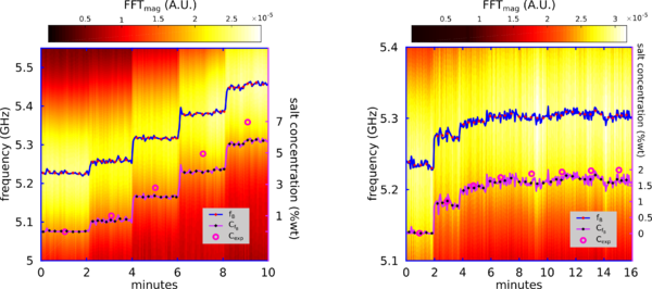
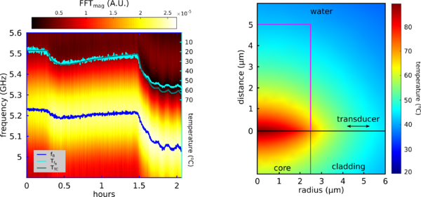
Viscoelastic properties of the probed medium can be obtained by evaluating the frequency and attenuation rate of the sampled phonon. Through Brillouin scattering, the phonon frequency is directly related to the sound velocity of the medium, and therefore its elasticity. Therefore, by measuring the Brillouin frequency of an object, one gains access to the unique elastic signature of the object-material. This system can then be used as a spectrometer, which is capable of detecting in real-time when the fibre-tip is in contact with specific liquds: water, glycerol, gelatin, blood plasma, methonal, etc. The fibre-spectrometer is also capable of detecting and characterising changes in the temperature and salinity of an object. Through thermo-optic, thermo-acoustic, salino-optic, and salino-acoustic effects, changes in temperature or salinity will modulate refractive index and sound velocity, producing a shift in the Brillouin frequency detected by the sensor.
Viscoelastic properties are promising biomarkers for health and development; the mechanical properties of cells, tissues, and their surrounding environments greatly influence important biological processes such as migration and differentiation which impact things like the progression of tumours and the lineage of stem cells. Typical methods for assessing these properties of biological tissues, such as atomic force microscopy, are often invasive and only offer inspection of superficial features. A device such as the phonon probe discussed here offers endoscopic implementation with the potential for non-contact operation. Furthermore, by using optical fibres, the device is inherently compatible with needles, catheters, and endoscopes used in clinical settings. Therefore, there is great promise for using the probe as an in vivo spectrometer for inspecting the elasticity of bio-liquids. Since GHz frequency ultrasound is used, the acoustic wavelength of ~300 nm would provide access to spectroscopy on the cellular scale. Current and future development of this device will focus on modifying the system for parallelised spectroscopy and 3D imaging with microscopic resolution.
Publications
Salvatore La Cavera, Fernando Pérez-Cota, Rafael Fuentes-Domínguez, Richard J. Smith, and Matt Clark, "Time resolved Brillouin fiber-spectrometer," Opt. Express 27, 25064-25071 (2019)
https://www.osapublishing.org/oe/fulltext.cfm?uri=oe-27-18-25064&id=416986


